Percutaneous Nephrolithotomy (PCNL) is the procedure of choice for large, bulky renal stones, especially those of dense consistency.
Standard treatments for routine kidney stones, such as shock wave lithotripsy, Ureteroscopy with laser lithotripsy, or endoscopic removal via mini-endoscope through the ureter, are effective in dealing with most stones. However, more definitive steps are required when confronted with large stones of 1-2 cm or more in size compounded by those of harsh chemical makeup. This is where PCNL comes in.
How Does PCNL Work?
PCNL allows direct contact with the stones with full-size endoscopes and lithotripsy (stone busting) devices. These devices can use mechanical pulses or controlled laser energy to break up kidney stones under direct visualization. Simultaneous removal of stone fragments achieves the most expedient and complete stone clearance.
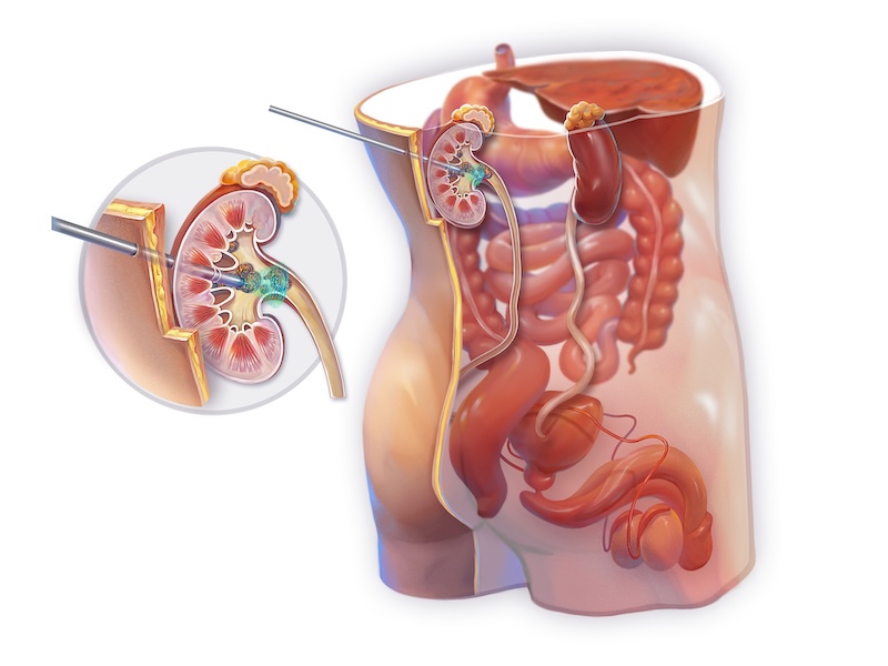
During PCNL, our radiology partners place single access or multiple accesses on the flank. Think of the kidney outlets as fingerlike. The finger structures allow access to different kidney areas and, thus, better access to the stone. Proper diagnosis and identification of the location of the kidney stone are critical to ensuring adequately placed accesses.
Once in place, the access is dilated to allow the placement of working ports, gaining entry into the kidney’s plumbing system where stones are. Various devices, including laser, ultrasound, or pneumatic jackhammer-like probes, are then used to disintegrate the stones. Sizable fragments are quickly evacuated without the need for the passage, which often is not feasible when dealing with a large amount of debris.
Example PCNL Surgery
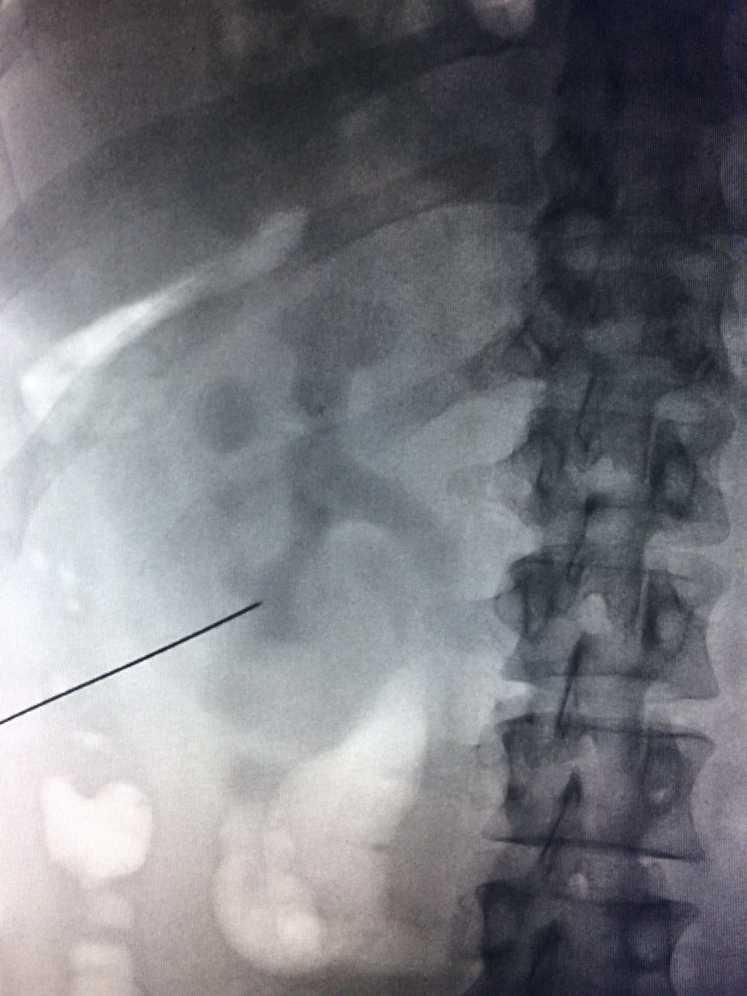
In this percutaneous nephrolithotomy, you will see Dr. Kemper visualizing a prominent kidney stone visible and accessible within the kidney. Dr. Kemper proceeds to break up the kidney stone into small pieces using a pneumatic-style kidney stone device. However, a second larger kidney stone is inaccessible via the existing access point. As a result, Dr. Kemper will have to perform the procedure again after new access is placed for the second stone. Had the second stone been accessible, Dr. Kemper planned on using laser lithotripsy to it up. In this case, the laser machine had been prepped and moved to the side of the table but could not be used because of access issues.
In addition to the vivid video from the scope within the kidney, continuous X-ray or fluoroscopy was also used to visualize the structures of the kidney and the stone itself. Dr. Kemper uses this fluoroscopy as his guidance tool in the first half of the procedure and typically does not require it for the rest of the procedure.
Patient Comfort During Surgery
After being prepped for surgery, the patient is wheeled into the operating room, still awake. At that point, the anesthesiologist uses a combination of IV anesthesia and laughing gas to put the patient to sleep for the duration of the procedure. The patient is placed face down on the operating table, with care not to disturb the previously placed access. Note: Typically, the access is placed in the flank one to two days before the surgical procedure.
A straightforward PCNL requires an overnight stay. Most patients are treated with a single session, but some require multiple sessions days apart to achieve a satisfactory result.
The Benefits of PCNL
As mentioned above, larger kidney stones lodged within the kidney are often best treated surgically with a PCNL. Fortunately, while surgery is daunting, kidney stone surgeons are very safe and effective in the hands of a qualified and experienced urologist who focuses on this treatment modality, like those at Georgia Urology.
Once in place, the access is then dilated to allow placement of working ports gaining entry into the plumbing system of the kidney where stones reside. Various devices including laser, ultrasound or pneumatic jackhammer like probes are then used to disintegrate the stones, sizable fragments are easily evacuated without the need for the passage which often is not feasible when dealing with a large amount of debris.
Videos Examples
The three videos below illustrate the large or staghorn stones we often encounter. They are named for their shape, as they take up the entire interior of the kidney.
Video 1: Before
Video 2: Multiple sessions in progress, with a marked reduction of stone burden and multiple drainage catheters in place.
Video 3: Post-op with minimum debris remaining.
Early management of kidney stones is crucial to effective treatment and avoidance of urinary issues, including kidney failure. Be sure to contact a urologist, such as those at Georgia Urology, to get an appointment for treatment and relieve the often-significant pain associated with stones. Our stone hotline is available 24 hours a day for appointments. We are often able to see stone patients the same or the next business day before the pain progresses.

 In this percutaneous nephrolithotomy, you will see Dr. Kemper visualizing a prominent kidney stone visible and accessible within the kidney. Dr. Kemper proceeds to break up the kidney stone into small pieces using a pneumatic-style kidney stone device. However, a second larger kidney stone is inaccessible via the existing access point. As a result, Dr. Kemper will have to perform the procedure again after new access is placed for the second stone. Had the second stone been accessible, Dr. Kemper planned on using laser lithotripsy to it up. In this case, the laser machine had been prepped and moved to the side of the table but could not be used because of access issues.